
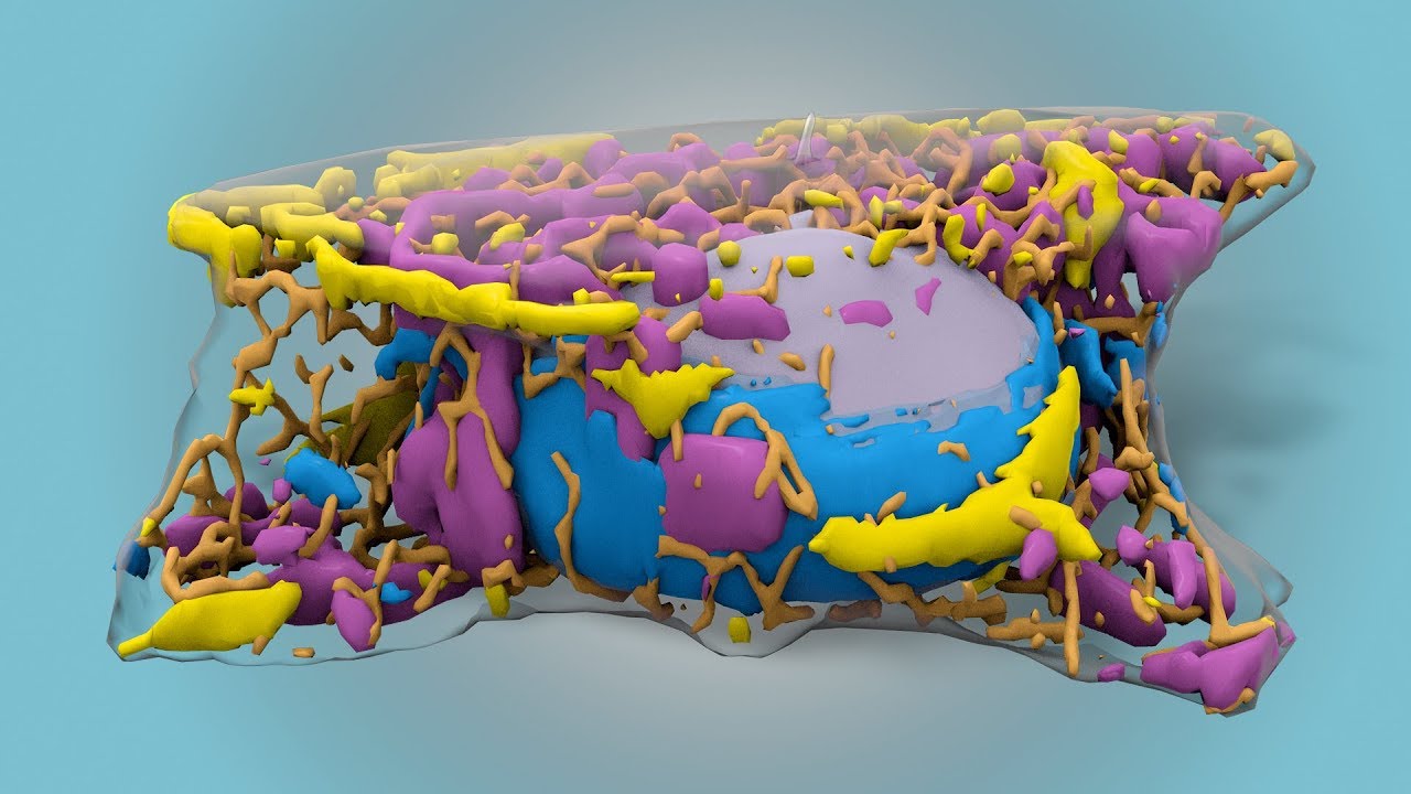
We all more than a year we hear about stem cells, but it can be seen is that in a digital microscope image which may not give the full picture. But a group of scientists from Allen Institute, using the deep algorithms of machine learning introduced an algorithm that is able to build a 3D model of a cell even if there is only a two-dimensional image of it.
In their design, the scientists taught the program to build the model cells, as it “highlight”. Thus all the structures and organelles become visible much better and also much more details. Typing several thousand photographs of these highlighted cells, scientists “fed” their neural networks. After that, the neural network was able on the basis of the image to predict the location of the organelles and their shape.
The experts of the Institute of Allen for several years engaged in similar developments, but in the past they were available only for internal use. The new program will be available to the public, and, according to the authors, will allow to understand the functioning of the cell and to observe all processes that occur within it. As noted by one of the authors Greg Johnson,
“If we will better understand the inner workings of healthy cells, we can understand what is happening, when it is modified. For example, turns into cancerous. This will give us the opportunity to see how cells change over time, we will be able to monitor all changes that occur and also to predict and detect them in vivo”.
#video | Scientists have shown how stem cell in 3D
Vladimir Kuznetsov