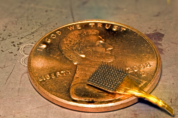
Eccentric in the best sense of the word entrepreneur, playboy, philanthropist Elon Musk known around the world. He decided to bring humanity into space, to colonize Mars, to abandon a disposable rockets. He decided to make the world cleaner, transplanted us from cars with internal combustion engines to self-driving cars. While unfolding these businesses, he does not sit idly by. He conceived Neuralink that will help us become new people. Without borders and without weaknesses, as it should be in the new world (Elon musk). To document the crazy ideas Mask, as always, volunteered Tim urban from WaitButWhy (he wrote about artificial intelligence, colonization of Mars and SpaceX). We present one of the best works of modern popular science journalism. Further, in the first person.
Part 1: Colossus Of Human
Part 2: Brain
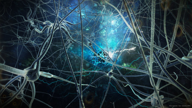
Flying over the nest of neurons

Letís go back in time to 50,000 BC, steal and somebody will bring it in 2017.

This Is The Side. Side, thank you and your men for what you’ve invented a language.

To thank you, we want to show you all the incredible things we were able to build thanks to your invention.

Okay, let’s put Boca on the plane, then a submarine, then pulled at the top of the Burj Khalifa. Now let’s show him a telescope, TV and iPhone. And let a little sit on the Internet.
It was fun. How do you Side?

Yes, we understand that you are very surprised. For dessert, let’s show him how we communicate with each other.


Side would be appalled if he knew that, despite all the magical abilities that people have acquired as a result of dialogues among themselves, thanks to the ability to speak, the process is no different from what was in his time. When two people are going to talk, they used technology of the age of 50 000 years.
Side also surprised that in a world in which work amazing machines, the people who did these cars go around with the same biological bodies, which went Sideways and his friends. How is this possible?

That’s why brain-computer interfaces (NCI) is a subset of the broader field of neural engineering, which itself is a subset of biotechnology, is so interesting. We have conquered the world with their technology, but when it comes to the brain is our main tool — the world of technology gives us nothing.
Therefore, we continue to communicate using technologies invented by Side. So I type this sentence 20 times slower than we think, and therefore, disease related to brain, still claim too many lives.
But after 50,000 years after the great discoveries the world can change. The next frontier brain he is.
* * *
There are many different options for potential brain-computer interfaces (sometimes referred to as the interface “brain — computer or brain — machine”) that are useful for different things. But everyone who is working on CQM, trying to solve one, two or both of these questions:
- How am I going to extract the desired information from the brain?
- I’ll send the desired information to the brain?
The first is the withdrawal of the brain — that is, recording what they say neurons. The second relates to the introduction of the information in the natural flow of the brain or change the natural flow in some way — that is, stimulation of neurons.
These two processes always occur in your head. Right now your eyes fulfil a set of horizontal movements that allow you to read this sentence. It is the neurons of the brain provide information to the machine (your eyes), and the car receives the command and responds. And when your eyes move a certain way, the photons from the screen to penetrate into your retina and stimulate the neurons in the occipital lobe of your cortex, allowing the picture of the world to get you in consciousness. Then this picture stimulates neurons in another part of your brain that allows you to process the information contained in the image, and to extract meaning from sentences.
The input and output of the information — that’s what makes brain neurons. The whole industry NRI wants to join this process.
At first it seems that this is not such a difficult task. Because the brain is just a ball of jelly. And the cortex — the part of the brain that we want to add to our recording and stimulation — it’s just a napkin, situated on the outer part of the brain, where it is easy to access. Inside the cortex has 20 billion neurons, 20 billion little transistors that can give us a completely new way to control our life, health and peace if we learn to work with them. Is it so hard to understand? The neurons are small, but we know how to split the atom. The diameter of the neuron in the 100 000 times more atoms. If the atom were a Lollipop, a neuron would be a kilometer in diameter — so we definitely should be able to work with such quantities. Right?
What is the problem?
On the one hand, it is correct idea because they lead to progress in the field. We really can do it. But once you start to understand what is actually happening in the brain, it becomes immediately obvious that this is the most difficult task for a person.
So before we talk about the CQM, we need to look at what people are doing are creating NKI. It is best to increase the brain 1000 times and see what happens.
Remember our comparison of the cerebral cortex with tissue?
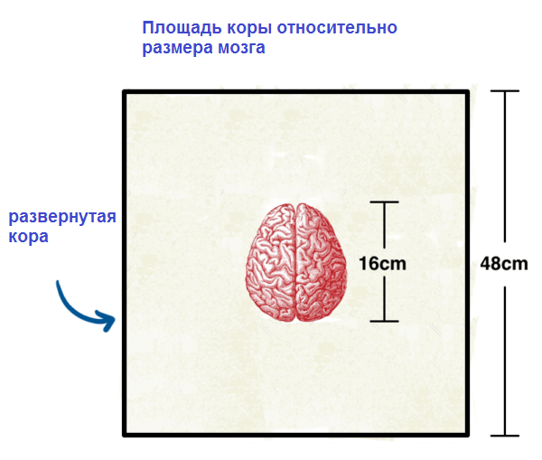
If we increase the tissue of the bark 1000 times — and she was about 48 inches on each side — now it will be the length of two blocks in Manhattan. It will take about 25 minutes to walk the perimeter. And the brain in General is the size of Madison Square garden.
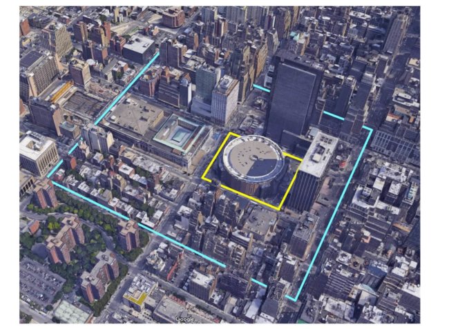
Let’s put it in the city. I’m sure several hundred thousand people who live there, we understand.

I chose 1000-fold increase for several reasons. One of them is that we all can instantly convert dimensions in your head. Every millimeter the actual brain became a meter. In the world of neurons, which is much less each micron became a millimeter, which is easy to imagine. Second, the bark becomes “human” dimensions: 2 mm thickness now 2 meter high person.
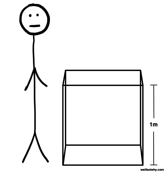
Thus, we can come to 29th street, to the edge of our giant swipe, and it’s easy to see what was going on in her two-meter thickness. To demonstrate, let’s pull out our giant cubic meter of bark, to explore it, to see what happens in a normal cubic millimeter of real bark.
What we see in this a cubic meter? Hash. Let’s clean it and put it back.
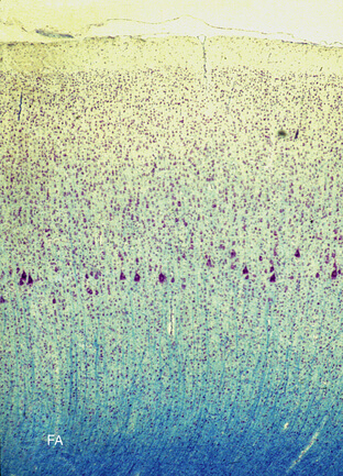
First place catfish, small bodies of the neurons who live in this cube.
Catfish vary in size, but the neuroscientists, with whom I spoke, say that the soma of neurons in the cortex, most often 10-15 microns in diameter (one micron = micron, 1/1000 of a millimeter). That is, if you put 7-10 of these in a line, this line will be the diameter of human hair. In our scale soma will be 1-1,5 cm in diameter. Lollipop.
The volume of the entire crust fits 500 000 cubic millimetres, and this space will be about 20 billion soms. That is, the average cubic millimeter of cortex contains about 40,000 neurons. In terms of our around 40 000 cubic meter of candy. If we divide our box into 40,000 blocks, each with the face 3 of an inch, each of our soma-candy will be at the center of its own 3-inch cube, and all other catfish — 3 inches in all directions.
Are you still here? Can you imagine our metre cube with 40,000 floating candy?
Here is a microscopic image of soma in the real cortex; everything else around it was removed:
Okay, so far it looks not so difficult. But soma is only a tiny part of each neuron. From each of our stretch Lollipop, twisted, branched dendrites, which in our scale can take three to four meters in different directions, and may be axon length of 100 meters (if transferred to another part of the crust), or kilometers (if descends in the spinal cord and the body). Each thickness in mm, and these wires make the crust in tightly twisted electrical vermicelli.
In this vermicelli is a lot of things. Each neuron has synaptic connections with a 1000 — sometimes up to 10 000 other neurons. As in the bark of about 20 billion neurons, this means that it will be more than 20 trillion individual neural connections (and a quadrillion connections in the brain). In our cubic meter is more than 20 million synapses.
With all this, not only from each Lollipop from 40 000 in our Cuba come a thicket of vermicelli, but thousands of other spaghetti pass through our cube from other parts of the cortex. And so, if we tried to record signals or to stimulate neurons specifically in this cubic region, we had to be very hard, because in the mess of spaghetti it will be difficult to determine which threads belong to our spaghetti soma-candy (and God forbid, in principle, be Purkinje cells).
And, of course, don’t forget about neuroplasticity. The voltage of each neuron changes constantly, hundreds of times per second. And tens of millions of synaptic connections in our cube will constantly change sizes, disappear and reappear again.
But this is only the beginning.
It turns out that in the brain there are also glial cells — cells that are of different types and perform many different functions, such as leaching of chemicals released in the synapses, wrap axons with myelin and maintenance of the immune system of the brain. Here are some of the most common types of glial cells:
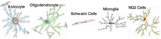
And how many glial cells are in the cortex? About the same as neurons. So add in our cube 40 000 of these.
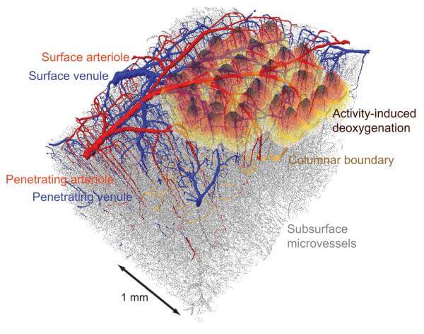
Finally, there are blood vessels. In each cubic millimeter of cortex contains about a meter tiny blood vessels. In our scale this means that in our cubic meter are the kilometer of blood vessels. Here’s how they look:
The retreat on the theme of connectomes
There is a lovely project I’m working neuroscientists, it’s called the connectome project human (Human Connectome Project). Scientists are trying to create a full detailed map of the entire human brain. Earlier anybody and close did that.
The project involves slicing the human brain for very thin plates is about 30 nanometers thick. It is 1/33, 000 millimeter.
In addition to creating great images “ribbon” formations of axons with similar functions which often develop within the white matter, like this —
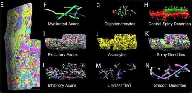
— the connectome project helps to visualize how all that stuff Packed in the brain. Here is a detailed breakdown of everything that happens in a tiny slice of mouse brain (blood vessels):
(For image, E — the complete section of the brain, and the F – N — the individual components that comprise the E).
So, our meter box is Packed, littered with electrified filling of different complexity. Let us now remember that in fact, our box — a cubic millimeter in size.
Engineers brain-computer interfaces need to either figure out what they say microscopic catfish, buried to the millimeter, or to stimulate certain soma that they do the right things. Good luck to them.
It would be difficult to do this with our enhanced 1000 times by the brain. With the brain, which well turns into a napkin. But actually he isn’t — this napkin is on top of the brain, highly folded (which, in our scale, depth 5 to 30 metres). In fact, less than a third swipe-crust is on the surface of the brain — most of it is in the folds.
In addition, the material with which you are able to work in a lab, not so much. The brain is covered by multiple layers, including the skull — which at 1000 magnification will be a 7-meter thick. And since most people don’t really like it when their skull is too long an open — and indeed it is questionable activity, they have to work with tiny lollipops of the brain as you can carefully and delicately.
And all this despite the fact that you are working with a bark — but a lot of interesting ideas on the subject of NKI are dealing with structures which are a lot below and if you stand on top of our city’s brain, they will lie at a depth of 50-100 meters.
Just imagine how much is going on in our cube but it’s just one 500 000 part of the cerebral cortex. If we smashed our giant bark in the same metre cubes and floated them in a row, they would stretch 500 kilometers to the Boston. And if you decide to make a bypass that will take more than 100 hours when walking fast at any moment you can stop and look at the cube, and all this complexity will be inside him. All this is now in your brain.
Neuralink Elon Musk. Part 3: how you should be happy if all that doesn’t bother you
Woweeeee.
Back to part 3: one flew over the nest of neurons
How do scientists and engineers will cope with this situation?
They try to make the most of the tools that they now have the tools that are used to record or stimulate neurons. Let’s examine the options.
Instruments NKI
With what has already been done, we can distinguish three broad criteria by which are evaluated the pros and cons of recording tool:
1) Scale — how many neurons can be recorded.
2) Resolution — how detailed the information gets the tool — spatial (how close your notes indicate which of the individual neurons are activated) and temporal (how well can you determine when there is recorded you activity).
3) Invasiveness — do I need surgery, and if so, how expensive.
Long-term goal is to skim the cream from all three and eat. But yet inevitably the question arises, which of these criteria (one or two) you can neglect? The choice of tool is not the increase or decrease of quality, is a compromise.
Let’s see what tools are currently in use:
fMRI
- Scale: large (shows information from around the brain)
- Resolution: from low to middle spatial, very low — temporary
- Invasiveness: non-invasive
fMRI is often used not in NKI, and as a classical instrument recording — gives you information about what is happening inside the brain.
fMRI uses MRI technology magnetic resonance imaging. Invented in the 1970’s, MRI has become the evolution of x-ray CT scan. Instead of x-rays, MRI uses a magnetic field (along with radio waves and other signals) to generate images of the body and brain. Like this:
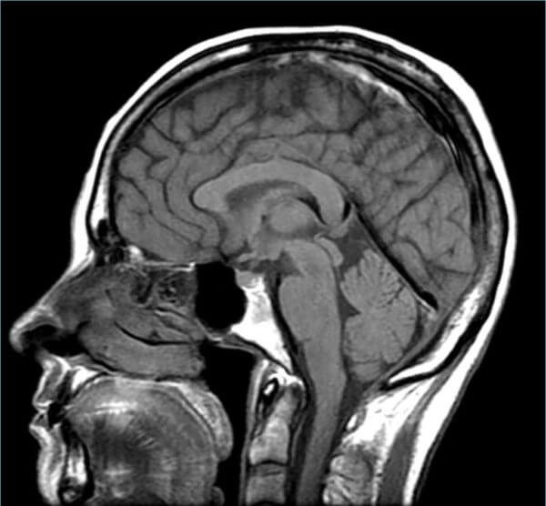
A complete set of cross sections that allows you to see the head as a whole.
Very unusual technology.
fMRI (functional MRI) technology uses MRI to track changes in blood flow. Why? Because when areas of the brain are more active, they consume more energy, and that means they need more oxygen so blood flow increases in this area to deliver this oxygen. Here’s what can show the fMRI scan:
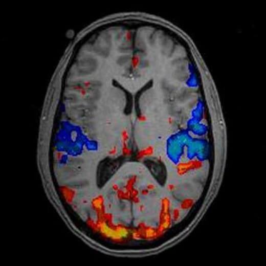
Of course, in the brain there is always blood — this image shows where increased blood flow (red, orange, yellow) and where it decreased (blue). And since fMRI can scan the whole brain, the results will be three-dimensional:
From fMRI many medical applications, for example, informing the physicians about how do certain parts of the brain after stroke, fMRI has learned very well from neuroscientists on what region of the brain involved in these functions. The scan also provides important information about what is happening in the brain at a specific moment in time, it is safe and non-invasive.
Biggest drawback is the resolution. an fMRI scan is a literal resolution as the computer screen pixels, but instead two-dimensional, the resolution is represented by three-dimensional cubic volumetric pixels — voxels (voxel, voxel).
Voxels fMRI has become smaller with the improvement of technology has led to an increase in spatial resolution. Modern fMRI voxels can be the size of a cubic millimeter. Brain volume is about 1 200 000 mm3, so scanning fMRI high resolution brain divides into one million small cubes. The problem is that at the neural scale is still quite a lot — each voxel contains thousands of neurons. So, at best, average fMRI shows blood flow, draw each group of 40,000 neurons or so.
Another big problem is the temporal resolution. fMRI monitors blood flow, which is inaccurate and is delayed about a second — an eternity in the world of neurons.
EEG
- Scale: high
- Resolution: very low spatial and medium-high temporary
- Invasiveness: non-invasive
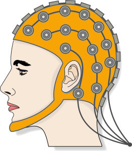
Invented almost a century ago, EEG (electroencephalography) imposes on the mind a lot of electrodes. Here it is:
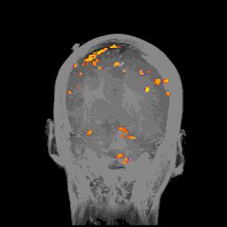
EEG is definitely a technology that will look funny for a primitive people in 2050, but at the moment it is one of the few tools that can be used with a completely non-invasive NKI. EEG records the electrical activity in different brain areas, displaying results as follows:
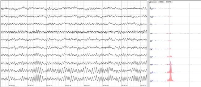
EEG graphs can reveal information on such medical problems as epilepsy, to monitor the sleep mode or to determine the condition of a dose of anesthesia.
Unlike fMRI, EEG has a fairly good temporal resolution, receiving electrical signals from the brain as they emerge — though the skull is greatly blurs temporal accuracy (the bone is a poor conductor).
The main drawback is the spatial resolution. The EEG it is not. Each electrode records only the mean value of the vector sum of the charges from millions or billions of neurons (blurred because of the skull).
Imagine that the brain is a baseball stadium, its neurons are the people in the crowd, and the information that we want to be instead of the electrical activity of the derivative of the vocal cords. In this case, EEG is the group of microphones outside the stadium, for its outer walls. You can hear the crowd start to chant and can even predict what she was screaming about. You can make out the distinctive signals if there is a close fight, someone will win. You might also make out if something unusual. That’s about it.
ECOG
- Scale: high
- Resolution: low spatial and high timing
- Invasiveness: present
ECOG (electrocorticography) is similar to EEG, since it also uses electrodes on the surface — only places them under the skull on the surface of the brain.
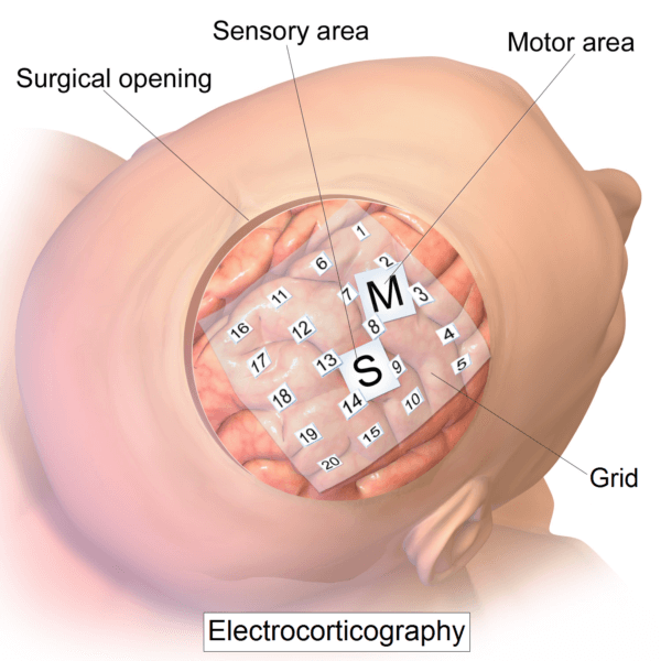
Dumb. But effective — much more effective than EEG. Without the interference that comes from the skull, ECOG covering over a high spatial (about 1 cm) and temporal resolution (5 milliseconds). The ECOG electrodes can be placed above or below the Dura mater:
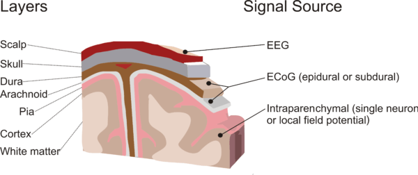
On the left the layers, top to bottom: scalp, skull, Dura, arachnoiditis, soft meninges, cortex, white matter. Right signal source: EEG, ECOG, intraparenchymally (LFP, etc.)
Returning to the analogy with our stadium, ECOG microphones are inside the stadium and closer to the crowd. So the sound will be much cleaner than microphones EEG outside the stadium, and ECOG will be able to distinguish the sounds of individual segments of the crowd. But is the improvement worth the money — requires invasive surgery. But according to the standards of invasive surgery, this intervention is not too bad. As I said one surgeon, “to put the stuffing under the Dura mater is relatively non-invasive. Will have to make a hole in the head, but it’s not so bad”.
The local field potential (LFP)
- Scale: small
- Resolution: medium-low spatial, high temporary
- Invasiveness: high
Let’s move with the surface of the electrode discs to the microelectrodes — tiny little needles that surgeons stick in the brain.
Brain surgeon Ben Rapoport told me how his father (a neuroscientist) did microelectrodes:
“When my father did the electrodes, he was doing them manually. He took a very fine wire — gold, platinum or iridium, which were 10-30 microns in diameter and inserted this wire into a glass capillary tube diameter in millimeters. Then held the glass over a flame and rotated until the glass is softened. He pulled a capillary tube until it becomes very thin and pulled out of the fire. Now capillary tube wraps and compresses the wire. Glass insulator, and the wire — guide. The result is an insulated glass electrode with a tip diameter of 10 microns”.
Although today some of the electrodes are still manufactured by hand, new technologies use silicon substrates and manufacturing techniques borrowed from the industry of integrated circuits.
The method of operation of the local field potentials is simple — you take one of those ultra-slim needle with an electrode tip and put it one or two millimeters in the cortex. There she collects the average value of the electric charges from all neurons within a certain radius of the electrode.
LFP provides you is not such a bad spatial resolution of fMRI combined with rapid temporal resolution of the ECOG. By the standards of the permissions is probably the best option of all the above.
Unfortunately, he’s terrible on other criteria.
Unlike fMRI, EEG and ECOG, LFP microelectrode has no scale — it only tells you what makes a small sphere surrounding it. And it is much more invasive as it actually enters the brain.
At the ballpark LFP is a single microphone hanging on one section with seats, removing sound clear in this area and maybe for a second or two snatches of an individual voice here and there — but for the most part he feels the overall vibration.
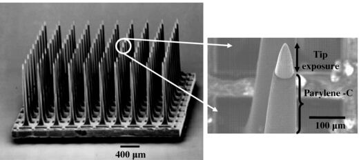
And my new development is a multi-electrode array, which is based on the idea of LFP that only contains 100 LFP at the same time. Multielectrode array looks like this:
A tiny square of 4 by 4 mm on 100 silicon electrodes on it. Here’s another one, here you can see how sharp electrodes is a few microns at the tip:
Registration of individual units
- Scale: tiny
- Resolution: superfine
- Invasiveness: very high
To record a wider LFP electrode tip a little kruglitsa to give the electrode a large surface area, and reduced resistance (an incorrect technical term) that one could detect some very weak signals from a wide range of locations. In the end, the electrode collects the chorus activity from the local field.
Check individual units also employ a needle electrode, but the tips are very sharp and make the resistance increase. Due to this displaced a large part of the noise and the electrode almost nothing picks up until you are very close to the neuron (about 50 microns), and the waveform of this neuron will be strong enough to overcome the wall of the electrode with high resistance. Obtaining separate signals from one neuron and not having background noise, this electrode can observe the personal life of the neuron. The smallest possible scale, the maximum possible resolution.
Some of the electrodes you want to bring relations to the next level and apply the local method of fixation potential (patch clamp), which allows you to remove the electrode tip and leave a tiny tube, glass pipette, which will directly suck the cell membrane of the neuron and carry out a more subtle dimension.
Patch clamp has the advantage: unlike all other methods, it physically touches the neuron and can not only record, but also to stimulate a neuron, injecting current or maintaining voltage at a certain level to perform specific tests (other methods can only stimulate groups of neurons as a whole).
Finally, the electrodes can conquer the neuron and actually penetrate through the membrane to record. If the tip is sharp enough, it will not destroy the cell membrane is like sealed around the electrode, and it will be very easy to stimulate neurons or to record the voltage difference between the external and internal environment of the neuron. But this is a short term technique — pierced neuron will not live long.
At our stadium, check the individual units will look like a unidirectional microphone attached to the collar of one fat man. Local fixation capacity is a microphone at someone in the throat, recording the exact movement of the vocal cords. This is a great way to learn about the experiences of the person about the game, but they will be taken out of context and it is impossible to judge what is happening in the game or about the man himself.
That’s all we have. At least that’s what we use quite often. These tools also very advanced and it will seem like stone age technology people of the future who do not believe that we had to choose one of the technologies to crack open the skull to obtain high-quality recordings about the brain.
But for all their limitations, these tools have taught us a lot about the brain and led to the creation of the first curious brain-computer interface. Read more about them in the next part.
To be continued.
Neuralink Elon Musk. Part three: flight over a nest of neurons
Ilya Hel