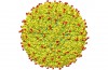

The above image is a 3D model of the virus zika, which became a real scourge for some countries in Central and South America. Now, thanks to the knowledge of the structure of the virus and its structure, scientists will be able to develop effective antiviral drugs and vaccines.
For the first time zika virus was discovered in Uganda in 1947 but at that time, scientists mistakenly thought it harmless. During the time of the outbreaks occurred in different places. However, the largest of these occurred in 2015, when the virus began a massive attack on the people of Brazil in April (currently 1.5 million confirmed cases of infection). By February 2016, the virus has covered 33 countries and territories on two continents. In this case, the predictions bleak: in their view, by the end of the year, the Zeke virus infecting 3-4 million people worldwide.
The virus is transmitted most often through mosquito bites. Main symptoms are fever, skin rash, conjunctivitis and pain in the joints. The sad part is that the virus leaves behind a very unpleasant consequences in the form of microcephaly (a condition in which children incorrectly develops the brain and as a consequence accompanied by a further cerebral disorders: autism, dementia and Guillain — Barre syndrome that can cause temporary paralysis) of children whose parents are ill with the virus during pregnancy.
Recent months, scientists are actively working to find a vaccine against this scourge and try to learn more about the virus. Recent successful research, a team of scientists from Purdue University became the first research group succeeded in visualizing the structure of zika virus. The details of this study wrote in the journal Science. Created almost atomic-level structural diagram of the virus will help scientists to find effective medicines.
“This discovery demonstrates not only the importance and significance of such fundamental research to human health, but also tries to draw attention to the rapidly increasing global problem”, said the rector of Purdue University Mitch Daniels.
“This talented group of researchers has solved one of the hardest and most important questions within a very short period of time and openly provided the results of its work for the further development of vaccines and therapies”.

Frozen cells of the virus zika
The leaders of the research group Richard Kahn and Michael Rossmann, and their colleagues created a three-dimensional map of Mature virus particles Zeke, using the technology of the so-called cryoelectron microscopy. After freezing the virus the scientists had sent him a stream of high energetic electrons created and thus a two-dimensional electron-microscopic images. Based on them, scientists have created a single three-dimensional model of the virus Zeke in high resolution. Usually in such cases, used x-ray crystallography, however, the new method has enabled much faster and more accurately to create the desired image.
The first thing the scientists noted was how the virus zika similar to other flavivirus — viruses transmitted through insect bites (e.g. dengue fever, West Nile, and dengue fever). Zeke, like other flavivirus, contains inside surrounded by a thick membrane icosahedral protein shell RNA genome. For scientists, this news is very happy, because it means that those techniques that were used to create vaccines against dengue and West Nile, can also be used to create a vaccine against the virus zika.

The Zeke virus in the context
And yet virus particles Zeke contain one very significant distinguishing it from other virus components. This component is the E-glycoprotein. It contains about 180 parts of this protein (marked in red), and so the virus is able to cling to certain human cells, including antibodies and cellular receptors. Similar to the “suckers” have dengue fever, but the unique features of the E-glycoprotein of the virus Zeke can explain how he is able to attack the nerve cells and also cells that play a key role in proper development of brain tissue.
“If these superficial “suckers” play the same role in dengue, allowing you to cling to healthy human cells, for us it is very good news, as it is a very important information base for the development of antiviral drugs,” said Rossmann.
“If all is true, it is quite possible to develop an inhibitor that can block the protein’s function of the virus to cling to healthy cells”.
In addition, it will be possible to develop a vaccine, which will focus on the destruction of the glycoprotein.